
Implantology and Oral Surgery

The Single and Reliable Choice for Your Health

Implantology and Oral Surgery
Dental Implants
A dental implant is an artificial tooth root surgically placed into the jawbone to replace a missing tooth. It is made from biocompatible materials such as titanium or zirconia. A crown, bridge, or prosthesis is then placed on top to restore chewing and aesthetic functions. Before treatment, a clinical examination and usually a three-dimensional tomography scan are performed to evaluate bone volume and density, allowing for surgical planning. The implant is placed under local anesthesia, and a period of generally 3–6 months is allowed for it to fuse with the bone (osseointegration); the duration may vary depending on bone quality and the implant site. Oral hygiene, smoking, diabetes control, general health, and teeth grinding habits are important factors in treatment success. With regular check-ups and proper care, implants can be used for many years.

Implantology and Oral Surgery
Surgical Implant
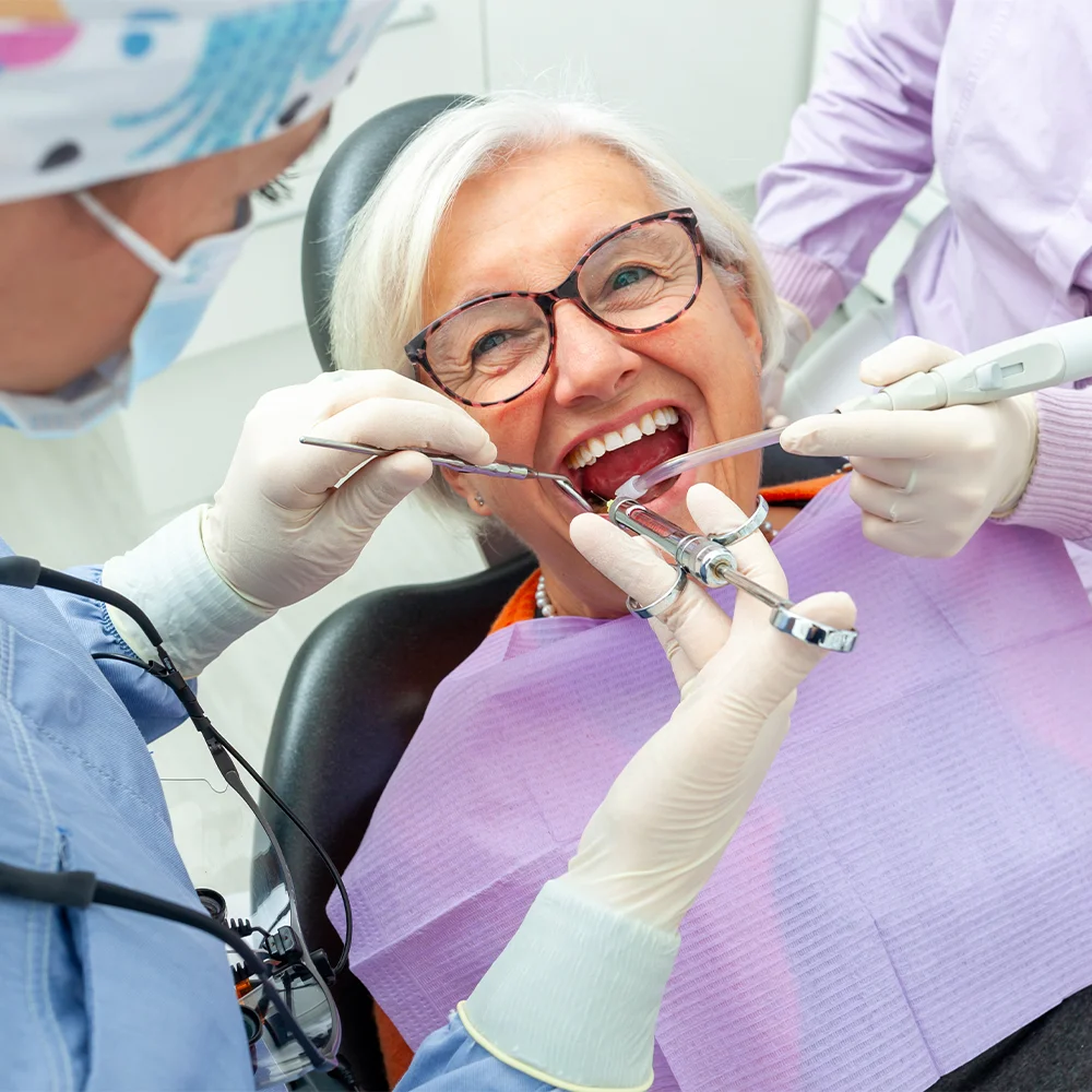
Surgical implant placement is a critical stage of treatment in which the dental implant is placed into the jawbone under precise and sterile conditions. Before the procedure, three-dimensional tomography is used to evaluate bone volume and density, as well as anatomical structures such as the nerve canal and sinuses. A surgical guide may be used when necessary. Under local anesthesia, the gum is opened, an implant site is prepared in the bone, and the implant is placed. Bone grafting may be required in cases where there is insufficient bone. The procedure may take between 30 minutes and several hours, depending on the number of implants and the characteristics of the case. Swelling, bruising, and tenderness may occur afterward. During the first few days, it is important to avoid hard and hot foods, refrain from smoking, and maintain good oral hygiene. Osseointegration is monitored through X-rays, and sutures are generally removed after 1–2 weeks.
Implantology and Oral Surgery
Sutureless (Flapless) Implant
A sutureless implant is a less invasive surgical technique in which the gum is not incised or lifted. Instead, the implant is placed directly into the bone through a small opening suited to the implant diameter. The implant’s position, angle, and depth are determined in advance using three-dimensional tomography and digital planning software; a surgical guide is generally used. Since there is no incision or suturing, post-operative swelling, bleeding, and discomfort may be less than with the conventional method. Recovery and return to daily activities may also be faster. This technique is preferred in suitable cases where there is sufficient bone volume and healthy gum tissue. Conventional surgery may be more appropriate in cases requiring bone grafting or involving complex anatomical structures. After the procedure, it is important to maintain good oral hygiene, avoid hard foods, and attend follow-up appointments.
Implantology and Oral Surgery
Ceramic Implant
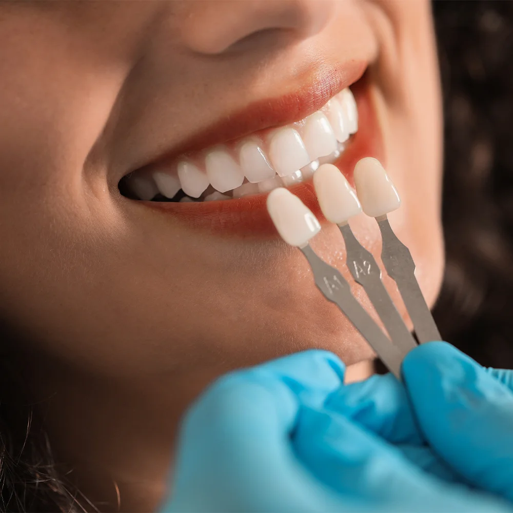
A ceramic implant is a metal-free, biocompatible type of implant made from zirconium dioxide, serving as an alternative to conventional titanium implants. It offers an option particularly for patients with metal sensitivity or allergies, or those who prefer a completely metal-free treatment. The white color of zirconium dioxide provides an aesthetic advantage by preventing the appearance of a grey metallic color, even if the implant neck becomes visible due to thin gums or gum recession. Its surface properties may help resist plaque accumulation. The placement process is similar to that of titanium implants: the bone structure is evaluated through imaging, and the implant is placed under local anesthesia. Its typically one-piece design may offer advantages in certain cases, while potentially limiting prosthetic flexibility. Ceramic implants also require regular check-ups and careful oral hygiene.
Implantology and Oral Surgery
Different Implant-Supported Prosthetic Options
Implant-supported prosthetic options are restorations determined according to the extent of tooth loss, bone condition, and functional expectations. In cases of a single missing tooth, a single crown secured to the implant with a screw or cement provides a result that closely resembles the appearance of a natural tooth. For multiple missing teeth, implant-supported bridges placed on two or three implants may be preferred. In cases of complete tooth loss, fixed systems such as All-on-Four or All-on-Six, secured onto four to six implants, or removable implant-supported dentures retained by attachments such as bars or locators may be used. The selection process takes into account the number of implants, bone density, budget, manual dexterity, aesthetic expectations, and ease of maintenance. Regular check-ups and proper care help support the long-term use of these prosthetic restorations.
Implantology and Oral Surgery
Bone Grafting
Bone grafting is a surgical procedure performed to address insufficient bone volume and density in the jawbone caused by tooth loss, long-term missing teeth, periodontal disease, or trauma. It can be performed before implant placement or, when necessary, during the same session as implant placement. Graft materials include autogenous grafts taken from the patient’s own bone, allogeneic grafts of human origin, xenogeneic grafts of animal origin, and synthetic alloplastic materials. The choice depends on the size of the defect and the clinical requirements. In cases involving larger grafts, a period of 4–9 months may be needed for the bone to mature. Sinus lifting may be performed when there is insufficient bone in the sinus area. Swelling, bruising, and tenderness may occur after the procedure. Avoiding smoking, maintaining good oral hygiene, and attending regular radiographic check-ups are important for graft healing and the success of implant treatment.
Implantology and Oral Surgery
Tooth Extraction
Tooth extraction is the removal of teeth that cannot be preserved, including those that are severely decayed, fractured, have lost their supporting structures due to advanced periodontal disease, or need to be removed to create space for orthodontic treatment. Before the procedure, an X-ray is used to evaluate the root structure, surrounding bone, and anatomical structures to determine whether the extraction will be simple or surgical. A simple extraction is performed under local anesthesia using elevators and forceps. Protecting the blood clot that forms after extraction is important for healing; spitting, using a straw, and smoking should be avoided during the first 24 hours. Mild bleeding, swelling, and tenderness may occur. Since leaving the gap for an extended period can lead to bone loss and shifting of adjacent teeth, options such as implants, bridges, or dentures should be planned in a timely manner. Healing is generally completed within 1–2 weeks.
Implantology and Oral Surgery
Impacted Tooth Extraction
Impacted tooth extraction is the surgical removal of teeth that remain completely or partially beneath the jawbone or gum tissue and have been unable to follow their normal eruption path. This may occur due to insufficient space in the jaw, obstruction by adjacent teeth, an abnormal angle of development, or genetic factors. It is particularly common with wisdom teeth and impacted canines. Before the procedure, a panoramic X-ray or three-dimensional tomography scan is used to evaluate the tooth’s position and its relationship with adjacent teeth and the nerve canal. Under local anesthesia, the gum tissue is accessed and, when necessary, some bone is removed. The tooth may be extracted in sections. The area is sutured after the procedure. Swelling, bruising, and tenderness may occur; cold compresses, prescribed medication, and a soft diet are recommended. Sutures are generally removed after one week.
Implantology and Oral Surgery
Impacted Wisdom Tooth Extraction
Treatment of impacted wisdom teeth involves the evaluation and, when necessary, removal of third molars that cannot fully erupt due to insufficient space in the jaw or that develop at an abnormal angle. In partially erupted teeth, the accumulation of food and bacteria can lead to pericoronitis, pain, swelling, and difficulty opening the jaw. Fully impacted teeth may cause pressure on adjacent teeth, root resorption, or cyst formation. The treatment decision is made by evaluating the tooth’s position, adjacent structures, the patient’s age, and symptoms. Teeth that do not cause problems may be monitored radiographically. When necessary, the tooth is removed using a simple or surgical technique; in surgical cases, bone removal and sectioning of the tooth may be required. Extractions performed at a younger age generally heal faster and result in fewer complications.
Implantology and Oral Surgery
Primary Tooth Extraction
Primary tooth extraction is the removal of primary teeth that are severely decayed and cannot be saved, have sustained serious damage due to trauma, or are preventing the eruption of permanent teeth. Since primary teeth serve as natural space maintainers for developing permanent teeth, as well as playing a role in nutrition and speech, a space maintainer appliance may be necessary after early tooth loss to prevent adjacent teeth from shifting. The decision to extract is planned by evaluating the extent of decay, root resorption, the position of the permanent tooth bud, and the child’s age, with X-rays taken when necessary. Extraction performed under local anesthesia is generally quick because the roots naturally resorb. To reduce the child’s anxiety, calm communication, play-based explanations, and, when necessary, mild sedation may be used. Mild bleeding and swelling may occur after the procedure; avoiding hard foods and not touching the area are recommended.